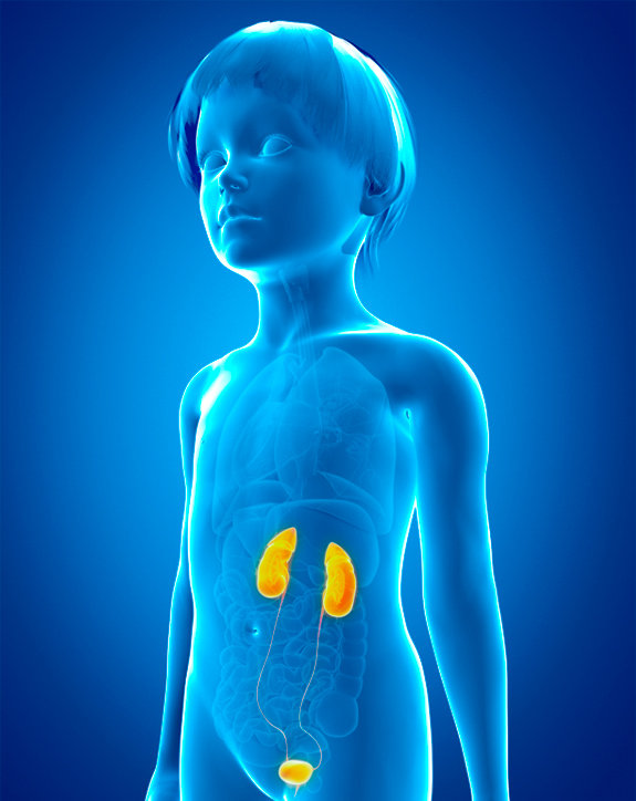
Compared to other parts of the world, the United States has the highest percentage of metabolic stone formers among the pediatric stone population. This is primarily related to hypercalciuria among pediatric stone formers in the United States.
Among children who are stone formers, there are multiple causes for stone formation, including renal tubular syndromes, cystinuria, , hypercalcemia, hypercalciuria, uric acid lithiasis, and enzyme disorders.
-
- Renal Tubular Disorders
- Cystinuria
- Hypercalcemia/Hypercalciuria
- Hypercalciuria
- Uric Acid Lithiasis
- Enzyme Defects
- Xanthinuria
- Hyperoxaluria
Renal Tubular Disorders
The two most common types of renal tubular disorders are renal tubular acidosis Type I (RTA) and cystinuria. Type I RTA is a disorder of hydrogen ion (H+) excretion, resulting from a tubular defect within the nephron which prevents a patient from generating a normal pH gradient between the blood and tubular urine pH. As a result, hyperchloremic acidosis is produced with excessive urinary losses of calcium, sodium, potassium, and phosphate. Nephrocalcinosis and calcium phosphate stone formation can occur as a result of hypercalciuria and low urinary citrate excretion.
The diagnosis results from the patient's inability to form an acidic urine (pH less than 5.5).
Treatment of Type I RTA is replacement of bicarbonate, sodium, and potassium with either sodium bicarbonate and potassium supplements or Polycitra K, which contains sodium citrate, potassium citrate and citric acid. The goal of treatment is to treat the hypocitraturia.
Cystinuria
Cystinuria is an autosomal recessively inherited disorder of tubular amino acid transport. This results in excessive urinary excretion of the basic amino acids, including cystine, ornithine, lysine, and arginine (C-O-L-A), and the formation of renal calculi. Normal individuals excrete less than 60 mg of cystine per 1.73 m2 of body surface area per day (1.73m2/per day). Patients who have this disorder have the excretion of cystine greater than 400 mg per 1.73m2/per day.
Cyanide nitroprusside test remains a widely used screening technique.
The treatment of cystinuria involves urinary alkalization and hydration. Medical therapy includes the use of D-Penicillamine, Thiola, or Captropil when stone formation reoccurs.
Hypercalcemia/Hypercalciuria
Hypercalcemia or increased levels of calcium in the blood can be associated with stone disease in children. The causes for hypercalcemia include:
-
- Primary Hyperparathyroidism
- Hypervitaminosis D
- Sarcoidosis
- Milk/Alkali Syndrome
- Neoplasia
- Cushing's Syndrome
- Hyperthyroidism
- Immobilization
Immobilization as a cause for stone disease occurs primarily in the pediatric population with head injuries, orthopedic injuries and severe burns. As a result of immobilization, there is increased bone reabsorption resulting in both serum and urine calcium levels to rise. The management of these patients should include adequate hydration and early ambulation, or calcitonin therapy if ambulation is not possible.
In the United States, Hypercalciuria appears to be a leading metabolic cause of stone disease accounting for 27-42% of pediatric patients with stone disease.
The etiology of hypercalciuria relates to 1) Absorptive Hypercalciuria, which is the hyperabsorption of intestinal calcium, or 2) Renal Hypercalciuria, which is the defective renal tubular absorption of calcium. Primary hyperabsorption of calcium from the gut increases serum levels of calcium, causing an increase of calcium in the urine. Treatment has been directed toward decreasing intestinal calcium absorption with neutral phosphate supplements and a low calcium diet.
Hypercalciuria
Renal hypercalciuria is characterized by the primary leakage of calcium from the kidney. The resulting mild hypocalcemia leads to an increase in calcium absorption in the gut. This condition is diagnosed by the presence of hypercalciuria in the fasting state. Thiazide diuretics and a low calcium diet are effective in reducing calcium excretion in patients with renal idiopathic hypercalciuria.
The current recommendation for determining the etiology of hypercalciuria is to obtain a 24-hour urine calcium excretion following one week of dietary calcium and sodium restriction. If urinary calcium remains elevated during calcium restriction, the patient is considered to have renal leak hypercalciuria. The correction of hypercalciuria by dietary restriction is consistent with the diagnosis of absorptive hypercalciuria. If a child has absorptive hypercalciuria, dietary calcium restriction to 400-600 milligrams per day is recommended. If these recommendations do not result in normalization of urine calcium excretion, hydrochlorothiazide (2mg/kg per day) is added. For a child with renal leak hypercalciuria that persists despite hydration and sodium restriction, hydrochlorothiazide (2mg/kg per day) is added.
A unique form of hypercalciuria related to stone disease has been recognized in neonates receiving furosemide therapy. It has become more prevalent with increasing numbers of neonates in intensive care settings who require furosemide for their lung disease. Furosemide results in hypercalciuria and calcium oxalate and calcium phosphate stone formation. The treatment of this disorder is medical and involves replacing furosemide with hydrochlorothiazide. In the majority of neonates managed medically, subsequent x-ray studies demonstrate a decrease or disappearance of renal calcifications.
Uric Acid Lithiasis
In the pediatric population, uric acid stones are seen primarily in children with 1) myeloproliferative or 2) intestinal tract disease.
Myeloproliferative diseases such as 1) leukemia and 2) lymphoma, typically lead to uric acid stone formation following courses of chemotherapy which result in increased purine turnover and excessive uric acid production.
Patients with intestinal tract diseases such as 1) regional enteritis and 2) ileostomy can experience excessive fluid and bicarbonate losses resulting in low volumes of acidic urine. This predisposes to uric acid crystal formation, which occurs at a pH of 5.7. Treatment of uric acid stone formation is medical. Alkalinization of the urine to pH equal to 6.5 and maintenance of a high urine volume results in stone dissolution in the majority of cases. Allopurinol may also be used in dissolving uric acid stones. Allopurinol works by inhibition of the enzyme xanthine oxidase, thereby decreasing the concentration of urinary uric acid.
Enzyme Defects
-
- Xanthinuria
- Hyperoxaluria
Xanthinuria
The enzyme defects xanthinuria and hyperoxaluria, are uncommon causes of pediatric stone formation. Xanthinuria is a rare, autosomal recessive disorder of purine metabolism caused by a deficiency of the enzyme xanthine oxidase, resulting in increased urinary xanthine and xanthine calculi, as well as hypouricemia and hypouricosuria. The most common cause of xanthinuria is the use of the medication allopurinol.
Hyperoxaluria
Primary hyperoxaluria is an uncommon autosomal recessive disorder of glyoxylic acid metabolism that results in excessive synthesis and excretion of oxalate. This disease results in urolithiasis or nephrocalcinosis before the age of 5. The diagnosis is confirmed by 24-hour urine excretion of oxalate in excess of the normal 40 milligrams per 24 hours.
Although infection stones are relatively uncommon in the United States, infection is the leading cause of pediatric stone disease in the United Kingdom, accounting for 2/3 of all cases. These stones are composed of magnesium ammonium phosphate, also referred to as struvite stones. The bacterium, Proteus mirabilis, is the most common organism in these types of stones.
Infection stones present earlier than other stone types. The male to female ratio is 2:1. The vast majority of infection stones were located in the upper urinary tracts (85%) and 15% were bilateral. Vesicoureteric reflux was diagnosed in 12% of children with infection stones.
The treatment of infection calculi include complete stone clearance and prevention of further infections. Nearly all patients with recurrent stones had reinfections. Therefore, it is important to correct any underlying cause for infection and keep these children on long term antibiotic prophylaxis.
Congenital anomalies of the urinary tract have been recognized as contributing factors of pediatric stone formation. 10-44% of children with stones are associated with congenital abnormalities of the urinary tract. Of the various congenital anomalies, ureteral pelvic junction obstruction was the most common factor seen in 54-65% of patients. Approximately 15% of anatomic stones were located in the lower urinary tract and were most commonly seen in children with bladder reconstruction or neuropathic bladder.
Treatment involves simultaneous correction of the obstructing abnormality at the time of surgical stone removal. In this group of patients, there is a high stone recurrence rate of approximately 27%.
Idiopathic stone formers comprise approximately 25% of pediatric stones. The syndrome of idiopathic calcium oxalate stone formation includes a group of heterogenous abnormalities for which the underlying causes are incompletely, or not well defined. This is a diagnosis of exclusion, made after other primary metabolic causes of stone formation have been eliminated. Seventy to 80 per cent of the patients who form stones in industrialized countries have this syndrome. In children, this percentage may not be this high, yet it remains the most common metabolic cause of stone formation within the urinary tract. Most often, it becomes symptomatic after the onset of puberty, and when patients with this syndrome begin to form stones before the age of 20, they usually demonstrate recurrent stone formation requiring specific treatment adjustments, including medication, to prevent further stone formation. This syndrome occurs commonly in families, and when it does, there is an autosomal dominant pattern of inheritance.
